
Type 1 is an sAVM of the dura (covering of the spinal cord) at the nerve root sleeve.

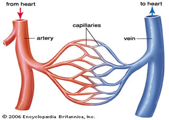
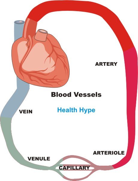
SAVMs were categorized in four groups by Anson and Spetzler in 1992. When an AVM occurs in the spinal cord or brain, the consequences can be significant. Used with permission of Mayo Foundation for Medical Education and Research, all rights reserved.ĪVMs can occur anywhere in the body. Surgical correction of a pediatric sAVM has lower consequences than with adults. This might be due to the elasticity of children’s blood vessels. SAVM in the pediatric population is rarer than in adults. Still others think they may be due to some neurological event like an undetected, small stroke. Current theories about AVM formation are that they are genetic or have a family tendency. At one time AVMs were thought to be a congenital abnormality meaning they occur during development of the fetus. The cause of a sAVM or other AVM is unknown. The risks for a second rupture of an AVM include when the AVM presents with hemorrhage, when there is deep venous drainage, when associated with an aneurysm, or when in a deep location. For example, you can have an sAVM bleed in the spinal cord where there is a significant nerve that is affected, or you can have a great deal of bleeding that affects many nerves in the spinal cord and extends into the brain. How your body is affected depends on the location of the bleed as well as the amount of bleeding that occurs. Nervous tissue damage creates functional deficits. Strokes can occur in the Central Nervous Systems which includes both the spinal cord and brain. Since sAVM in the spinal cord or AVM in the brain is a hemorrhage (bleeding) into tissue it is considered a type of hemorrhagic stroke. Due to the pressure of the extra blood pressing on the soft neurological tissue, a change in blood pressure at the site and bleeding into the surrounding neurological tissue results in damage to the spinal cord or brain.Īn AVM in the spinal cord is designated as a spinal AVM. The extra blood in the CNS will push on healthy nerve tissue depriving the spinal cord or brain of oxygenated blood flow. The brain is surrounded by the skull that also does not allow for expansion. If an AVM in the spinal cord or brain ruptures, there is no room for even a small amount of extra blood because the spinal cord is surrounded by bony vertebrae that do not expand. Some AVMs rupture in parts of the body that can accommodate some extra fluid in the tissue until it is absorbed.Īn AVM in the Central Nervous System (CNS) is a serious condition. In many cases, an AVM might be present but never ruptures. The result of the AVM is bleeding into tissue of the body.ĪVMs can occur in any part of the body. Over time, the walls of the blood vessels become thin and weak, leading to possible rupture. The area of the misconnection causes the blood vessel to dilate due to rapid blood flow through the artery which is not slowed by the capillaries. An AVM is often described as a ‘tangled web’ of vessels at the location of miscommunicated blood flow. This missed step results in an arteriovenous (artery to vein) malformation (AVM). Very rarely, an artery may connect directly to a vein bypassing the capillaries which are missing. Veins carry blood and waste products away from the cells. Body cells obtain the enriched blood through capillaries. In the body’s circulation, arteries deliver enriched blood to the cells. Donate to advance SCI and paralysis research.The North American Clinical Trials Network (NACTN).International Spinal Research Trust (ISRT).Developing Spinal Cord Injury Treatments.Tomorrow’s Cure - SCI and Paralysis Research.Ask Us Anything / Connect with an Information Specialist.Today's Care: National Paralysis Resource Center.


 0 kommentar(er)
